

Minnie's Sinus Mystery
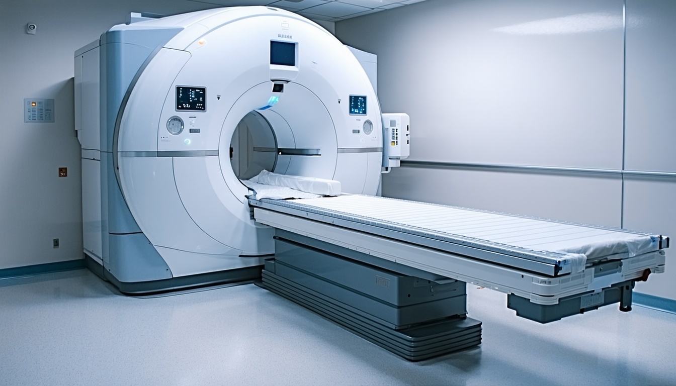
Veterinary medicine has undergone a remarkable transformation in recent decades, with advanced imaging technologies leading the charge in improving diagnostic accuracy and patient outcomes. Among these innovations, Magnetic Resonance Imaging (MRI) is emerging as a go-to modality for diagnosing many complex conditions in companion animals, offering unprecedented detail and diagnostic capabilities that are reshaping how veterinarians approach challenging cases. With the improved sensitivity for pathology MRI is challenging the old adage "CT for bone, MRI for soft tissue".
Why Choose MRI
MRI's superiority lies in its ability to provide exceptional soft tissue contrast and multi-planar imaging capabilities without ionizing radiation. Unlike traditional imaging modalities, MRI can differentiate between various soft tissue types with remarkable precision, making it invaluable for diagnosing conditions that were previously difficult or impossible to detect accurately.
The technology's non-invasive nature, combined with its ability to image in multiple planes (sagittal, transverse, and dorsal), provides veterinarians with a three-dimensional understanding of anatomical structures and pathological processes. This comprehensive visualization capability has made MRI indispensable for neurological, orthopedic, and soft tissue evaluations.
The Broad Spectrum of Conditions MRI Can Diagnose
 Neurological Conditions
Brain Tumors and Masses MRI excels at identifying and characterizing brain tumors, including meningiomas, gliomas, and pituitary adenomas. The technology can distinguish between tumor types, assess their relationship to surrounding structures, and evaluate the extent of cerebral edema or mass effect.
Spinal Cord Disorders Intervertebral disc disease, spinal cord tumors, and inflammatory conditions like meningomyelitis are precisely visualized through MRI. The technology can identify the exact location and severity of spinal cord compression, crucial information for surgical planning.
Seizure Disorders MRI can reveal structural abnormalities causing seizures, including cortical dysplasia, hippocampal sclerosis, and subtle brain malformations that other imaging modalities might miss.
Orthopedic Applications
Joint Pathology MRI provides detailed visualization of cartilage, ligaments, tendons, and synovial structures. It can detect early degenerative joint disease, cruciate ligament tears, and meniscal injuries with exceptional accuracy.
Soft Tissue Tumors MRI excels at characterizing soft tissue masses, determining their origin, extent, and relationship to surrounding structures, which is crucial for surgical planning and prognosis.
Bone Marrow Disorders MRI can detect bone marrow edema, infection, and infiltrative processes that may not be visible on radiographs until significant bone destruction has occurred.
Abdominal and Thoracic Imaging
Liver Disease MRI can identify hepatic masses, assess liver architecture, and detect early fibrotic changes that might not be apparent with other imaging modalities.
Cardiac Conditions Cardiac MRI can evaluate myocardial structure, function, and perfusion, providing detailed information about congenital heart disease and cardiomyopathies.
Conclusion
The ability to identify cases that would benefit from MRI imaging - before exhausting other diagnostic modalities - can dramatically reduce diagnostic timelines and improve patient outcomes. This growing understanding of when MRI can reveal what other modalities cannot is elevating the standard of care across veterinary medicine. The integration of MRI into diagnostic decision-making represents the natural evolution of veterinary practice toward more precise, efficient, and effective patient care.
