
MRI Machine
Patient: Casey, a 7-year-old Golden Retriever
Presenting Problem:
Casey came in with a two-month history of worsening hind limb weakness and difficulty climbing stairs. His owners also noticed he was reluctant to jump into the car, which was unusual for him.
Initial Diagnostics:
Casey’s veterinarian did a full physical exam and routine X-rays, which didn’t show any clear bone abnormalities. A CT scan was then performed to check for possible intervertebral disc disease or bony tumors. The CT came back inconclusive—while it showed mild disc protrusion at L2-L3, it didn’t fully explain Casey’s worsening weakness or pain response when his lower back was palpated.

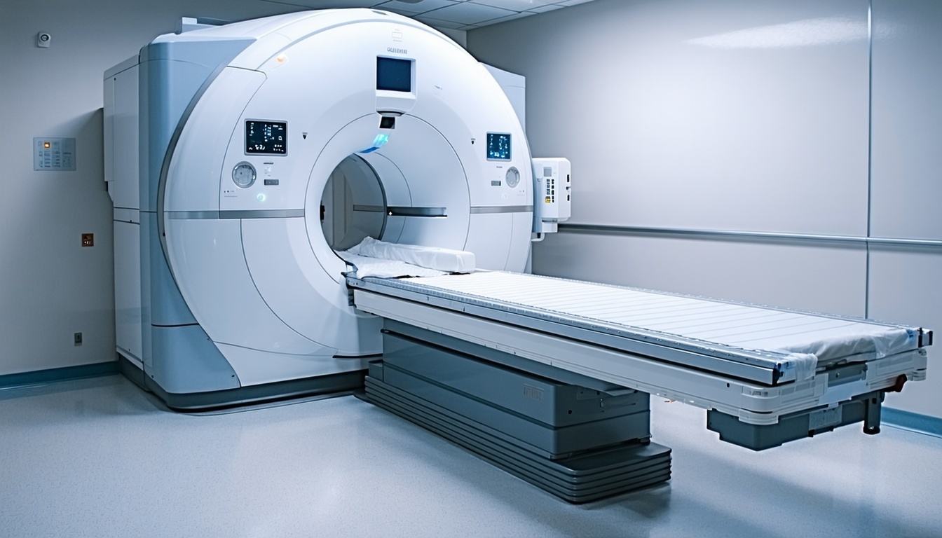
Next Step: MRI
Since his symptoms didn’t match the CT results, the vet referred Casey for an MRI. The MRI provided a detailed look at the soft tissues, spinal cord, and nerve roots L7S1 rupture with osteoarthritis suggesting lumbosacral instability. There was indeed a disc at L2-3 without compression of the thecal sac or cord edema. MRI interpretation was that this was a clinically insignificant disc and that his issue was at the lumbosacral space.
Outcome:
Casey was referred for decompression and stabilization surgery. He responded well and after 6 weeks was able to run and play normally.
Key Takeaway:
This case highlights how MRI can detect soft tissue and spinal cord problems that a CT scan might miss. For complex cases involving nerve or spinal cord compression, MRI often gives the detail needed to pinpoint the problem and guide proper treatment.
