
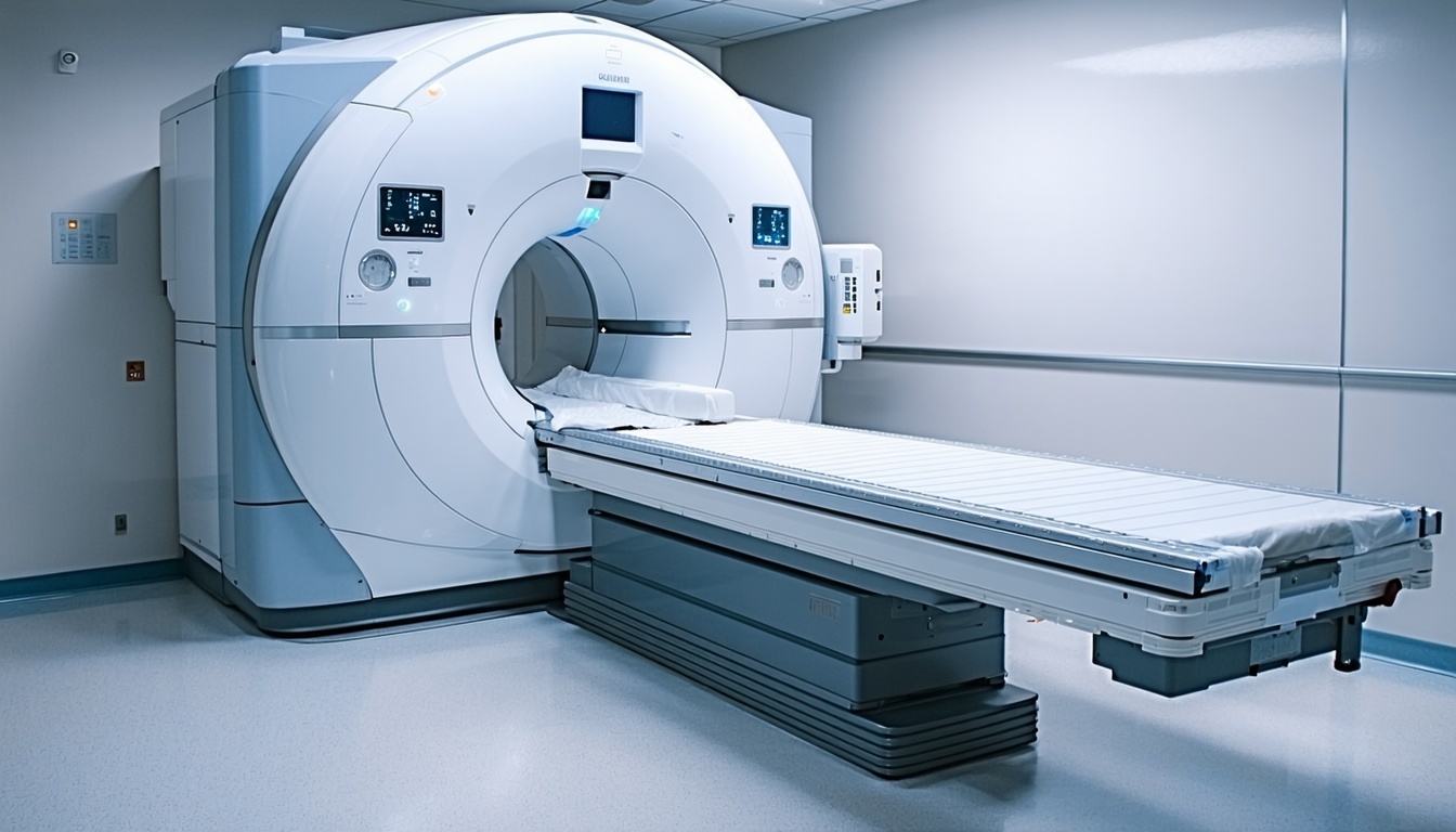
MRI Machine

Patient: Bella, a 9-year-old Labrador Retriever
Presenting Problem:
Bella had been losing weight for several weeks, seemed more tired than usual, and occasionally vomited after meals. Her blood work showed slightly elevated liver enzymes, so her vet suspected a liver issue.
Initial Diagnostics:
Abdominal ultrasound revealed an indistinct mass near the right kidney. The origin of the mass could not be determined, and fine needle aspirates were non diagnostic. Differentials included adrenal mass, renal mass, and retroperitoneal hemangiosarcoma or soft tissue sarcoma.
 Next Step: MRI
With the very disparate prognosis for the differentials Bella was referred for an MRI. MRI findings confirmed a right adrenal tumor without invasion into the vena cava. The enhanced soft tissue sensitivity and differing signal intensities for each of these tumor types allowed the distinction. Subsequent testing confirmed an adrenocortical tumor.
Outcome:
Because the MRI showed the mass’s exact location and its relationship to blood vessels, the surgical team could plan a precise liver lobectomy. The tumor was successfully removed and Bella continues to do well.
Key Takeaway:
This case shows how MRI can provide excellent soft tissue detail inside the abdomen, giving vets critical information when ultrasound and CT aren’t clear enough. In Bella’s case, MRI made the difference between uncertainty and a clear diagnosis that led to successful surgery.
